Conception is not a moment. It is a sequence of events that unfolds over hours and days, each step dependent on precise molecular timing. The process begins with two cells — a sperm and an egg — each carrying half the genetic material needed to build a human. Their meeting sets off a cascade that transforms a single fertilized cell into a structure that, within a week, begins to embed itself in the uterine wall and send chemical signals to the mother's body.
The sperm's journey: selection before fertilization
Of the 100 to 300 million sperm deposited during ejaculation, only a few hundred reach the site of fertilization in the ampulla of the fallopian tube. The rest are eliminated by vaginal acidity, cervical mucus, and the immune cells that patrol the female reproductive tract. The cervix acts as the first filter. Outside the fertile window, cervical mucus forms a dense mesh that sperm cannot penetrate. During ovulation, estrogen alters the mucus structure — it becomes watery and arranged in microscopic channels that guide sperm upward.
Sperm that pass the cervix undergo capacitation, a biochemical process that strips proteins and cholesterol from the sperm head membrane. Capacitation takes about 6 to 8 hours. Without it, sperm cannot penetrate the egg's outer layers. The process also primes the sperm for the acrosome reaction — the release of enzymes that digest the egg's protective coating.
The egg's preparation: a cell in waiting
The egg, or oocyte, is the largest cell in the human body — about 0.1 millimeter in diameter, just visible to the naked eye. It is arrested in metaphase II of meiosis, a state of suspended division it has maintained since before birth. The egg is surrounded by two barriers: the cumulus oophorus, a cloud of nourishing cells, and the zona pellucida, a tough glycoprotein shell.
At ovulation, the fimbriae of the fallopian tube sweep the egg inside. Cilia lining the tube create a gentle current that moves the egg toward the uterus. The egg is viable for about 12 to 24 hours. Fertilization must occur within this window.
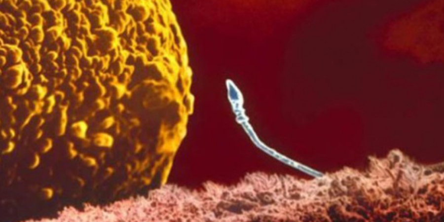
Fertilization: the moment of fusion
When sperm reach the egg, they encounter the cumulus cells first. Hyaluronidase enzymes released from the sperm acrosome dissolve the matrix holding these cells together. The sperm push through, reaching the zona pellucida. One sperm makes contact. Its acrosome ruptures completely, releasing enzymes that create a path through the zona. The sperm head fuses with the egg membrane.
Fusion triggers two immediate changes. First, the egg completes meiosis II, discarding half its chromosomes into a small polar body. Second, cortical granules inside the egg release their contents into the space between membrane and zona. This blocks additional sperm from entering — the zona reaction. Polyspermy, or fertilization by multiple sperm, produces a non-viable embryo with an abnormal number of chromosomes.
Inside the egg, the sperm tail dissolves. The sperm nucleus swells. Both nuclei — now called pronuclei — migrate toward each other. Their membranes break down. Maternal and paternal chromosomes mingle. This is syngamy, the formation of the zygote. The event takes about 18 to 24 hours after sperm entry. The cell now contains 46 chromosomes in a new, unique combination. Half from the mother, half from the father. The genetic blueprint is set.
Cleavage: the first divisions
About 24 hours after fertilization, the zygote divides for the first time. This is cleavage — mitosis without cell growth. The one cell becomes two, then four, then eight. Each daughter cell, called a blastomere, is smaller than the parent. The embryo remains the same overall size it was as a zygote, still enclosed within the zona pellucida.
At the 8-cell stage, around day 3, a critical change occurs: compaction. Blastomeres flatten against each other and form tight junctions. The embryo becomes a morula, Latin for "mulberry." Cells on the outside will later form the placenta. Cells on the inside will become the embryo proper. This is the first differentiation event in human development — the first decision about which cells will build the body and which will support it.
The blastocyst: a structure with a purpose
By day 5, fluid begins to accumulate inside the morula. The cells organize into a hollow ball called the blastocyst. It contains two cell populations:
- The trophectoderm. A single layer of cells forming the outer shell. It will give rise to the placenta and fetal membranes.
- The inner cell mass. A cluster of cells attached to one side of the cavity. These are pluripotent stem cells capable of forming any tissue in the human body. They will generate the embryo and, eventually, the entire organism.
The zona pellucida thins and ruptures. The blastocyst hatches. It is now free to interact directly with the endometrium, the uterine lining prepared by progesterone during the luteal phase. Hatching occurs around day 6. Implantation follows within 24 to 48 hours.
Implantation: the embryo embeds itself
Implantation is an active, invasive process. The blastocyst does not float passively into the uterine wall. Its trophectoderm cells extend protrusions that grip the endometrial surface. They secrete enzymes that dissolve the extracellular matrix, burrowing into the tissue. The embryo literally digs itself in. By day 9, it is fully embedded below the endometrial surface.
"The first seven days of human development remain one of the most difficult biological processes to study — not because it is unknowable, but because it happens in a place researchers cannot easily access without disrupting the very thing they want to observe." — Magdalena Zernicka-Goetz, developmental biologist, University of Cambridge
Once embedded, trophectoderm cells differentiate further into syncytiotrophoblast — a multinucleated mass that invades deeper into the endometrium and breaks maternal capillaries. Blood from these vessels fills spaces called lacunae, bathing the embryonic tissue. This is the beginning of placental circulation. The syncytiotrophoblast also produces human chorionic gonadotropin (hCG), the hormone detected by pregnancy tests. hCG signals the corpus luteum in the ovary to continue producing progesterone, preventing menstruation. This is the first chemical conversation between embryo and mother.
Gastrulation: the body plan emerges
Around day 14, the inner cell mass undergoes gastrulation — the defining event of early embryogenesis. Cells migrate and organize into three germ layers:
- Ectoderm. The outer layer. It will form skin, hair, nails, and the entire nervous system — brain, spinal cord, nerves.
- Mesoderm. The middle layer. It builds muscle, bone, blood, kidneys, gonads, and the cardiovascular system.
- Endoderm. The inner layer. It becomes the lining of the gut, lungs, liver, pancreas, and thyroid.
At this stage, a structure called the primitive streak appears — a line of cells that defines the head-to-tail axis of the future body. Gastrulation completes by the end of the third week. The embryo is now about 2 millimeters long but already organized along the fundamental axes that will persist for life.
From embryo to fetus: organogenesis
Weeks 3 through 8 constitute the embryonic period, during which every organ system forms. This is the most vulnerable phase of development. By day 22, a tube of cells begins to fold into a heart, and spontaneous contractions start. By day 28, the neural tube closes — the precursor to the brain and spinal cord. Failure of closure causes neural tube defects such as spina bifida, which is why folic acid supplementation is critical in the first weeks of pregnancy, often before a person knows they are pregnant.
By week 6, limb buds appear. By week 8, all major organs are present in rudimentary form. The embryo is about 3 centimeters long. After week 10, the term changes from embryo to fetus. The distinction marks the end of organogenesis. The remainder of gestation — roughly 30 weeks — is dedicated to growth, refinement of organ function, and the accumulation of adipose tissue.
A timeline of the first 28 days
The earliest stages of human life are rarely discussed in specific terms, yet they follow a reproducible sequence that embryology textbooks have documented for decades. The timeline below summarizes what happens from fertilization to the establishment of pregnancy:
- Day 0. Fertilization. Sperm fuses with egg. Pronuclei form.
- Day 1. First cleavage. Zygote divides into two cells.
- Day 3. Morula stage. 8–16 cells. Compaction begins.
- Day 5. Blastocyst forms. Inner cell mass and trophectoderm are distinct.
- Day 6–7. Hatching and implantation begin.
- Day 9–10. Implantation completes. hCG enters maternal blood.
- Day 14. Primitive streak appears. Gastrulation starts.
- Day 21. Heart begins beating.
- Day 28. Neural tube closes. Basic body plan established.
Every person alive has passed through this sequence. The process is ancient, shared across mammals with only minor variations. The molecules that drive it — the genes that pattern the body axis, the proteins that guide cell migration — are highly conserved through evolution. A mouse embryo and a human embryo look nearly identical during gastrulation, revealing the shared heritage written into the genetic code.
Development does not stop revealing itself. Each year brings new data from embryology labs and time-lapse imaging of in vitro fertilization procedures. The more we observe, the clearer it becomes that the beginning of life is not a single event but a progression — a series of carefully timed decisions made by cells that have no brain, no blueprint, only chemistry and physics shaped by a billion years of evolution.